
Strive to Improve Medical Conditions, Reduce Health Care Costs
Strive to Improve Medical Conditions, Reduce Health Care Costs
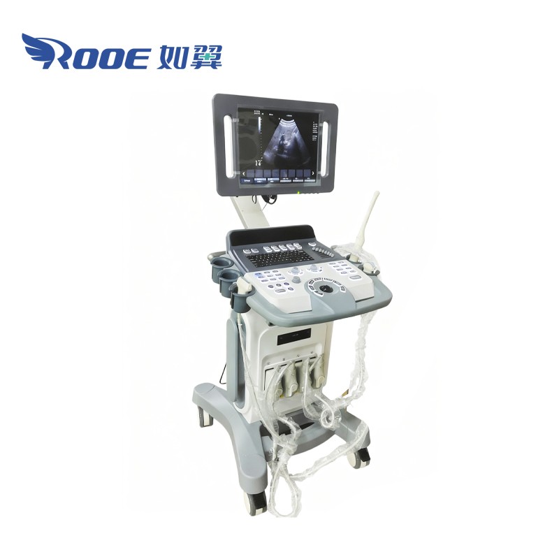

This is a high-performance color Doppler ultrasound diagnostic equipment designed for precise medical imaging and accurate diagnosis. Featuring advanced full digital technology, multiple probe options, and a high-resolution LCD display, it delivers clear, detailed images for various clinical applications including abdominal, cardiac, and obstetric examinations. This ultrasound imaging system offers stable performance, ergonomic design, and comprehensive connectivity, making it an ideal solution for hospitals, clinics, and medical diagnostic centers seeking reliability and efficiency in patient care.
This is a high-performance Color Doppler diagnostic device engineered to provide exceptional image clarity, operational efficiency, and reliable clinical results. Designed for hospitals, clinics, and medical imaging centers, it integrates advanced digital processing technology, a high-resolution medical LCD display, and multiple probe options to meet diverse diagnostic requirements with precision and consistency.
 |  |
1. Multiple Probe Options and Upgrades
Supports convex, linear, cardiac, transvaginal, and 4D probes, with optional upgrades for continuous Doppler and trapezoidal imaging.
2. High-Resolution Display
Equipped with a medical-grade LCD monitor and adjustable arm for optimal viewing.
3. Ergonomic Control Panel
Floating, backlit control panel ensures smooth operation under any lighting conditions.
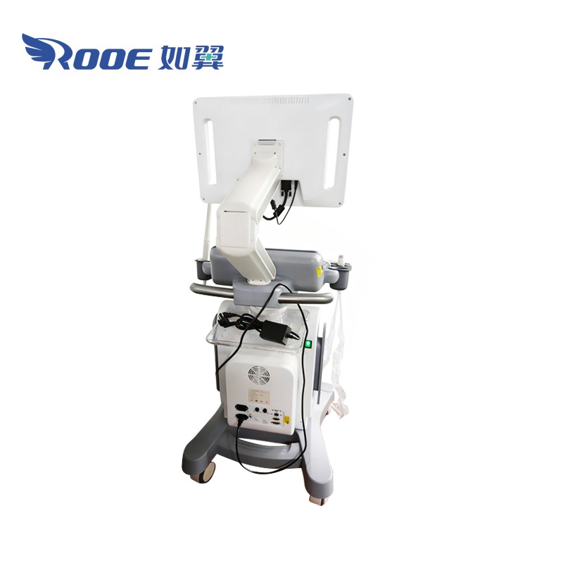
4. Multi-Port Connectivity
Four probe ports with USB and external interfaces for easy data transfer and printer connection.
5. Advanced Imaging and Doppler Functions
Full digital ultrasound system provides clear 2D and color Doppler images for accurate diagnostics.
6. PC-Based Platform with Large Storage
Built-in 1TB hard drive and open PC architecture support efficient data management and device compatibility.
7. Compact and User-Friendly Design
Lightweight (approx. 65 kg) with a multilingual interface, making it easy to use and move between departments.
 |  |
Probe configuration:
| Configuration | Probes | 5 Steps Multi-frequency |
| Standard | 3.5Mhz abdominal probe | 2.0, 3.0, 3.5, 4.0, 5.5Mhz |
| 7.5Mhz linear probe | 6.0, 6.5, 7.5, 10.0, 12.0Mhz | |
| Options | 6.5Mhz transvaginal probe | 5.0, 6.0, 6.5, 7.5, 9.0Mhz |
| 3.5MHz micro-convex probe | 2.0, 2.5, 3.5, 4.5, 5.0Mhz | |
| 3.5Mhz phased array probe | 2.1, 3.0, 3.5, 4.0, 5.5Mhz | |
| 3.5Mhz 4D volume probe | 2.0, 3.0, 3.5, 4.0, 5.5Mhz |
Technical Parameters:
| Displaying mode | B,B/B,4B,B/M,M, B/C,B/C/D,B/D, duplex, triplex, CFM, PW,CW(optional), 4D(optional) |
| Signal processing | Full-digital beam forming, dynamic filter, dynamic real time receiving focusing, RDA, DRA, spectral processing, CFM processing, real-time dynamic focusing, dynamic aperture in all fields |
| Image processing | THI HPRF Speckle-reduction Power adjustable Smoothing function Image optimization Tissue Harmonic imaging Dynamic apodization Dynamic aperture Edit the exam type and save the user‐defined items Pulse Wave Doppler Edge enhancement One-key optimization Image conversion Doppler Sound output volume adjustable Wall filter adjustable Base line adjustable Sampling frame adjustable Continuous Wave Doppler (Option) Trapezoid Image (Option) |
| Scanning depth | ≥300mm |
| Probe elements | 128 |
Strive to Improve Medical Conditions, Reduce Health Care Costs
© 2018-2020 Jiangsu Rooe Medical Technology Co., Ltd. All rights reserved. Site Map