
Strive to Improve Medical Conditions, Reduce Health Care Costs
Strive to Improve Medical Conditions, Reduce Health Care Costs

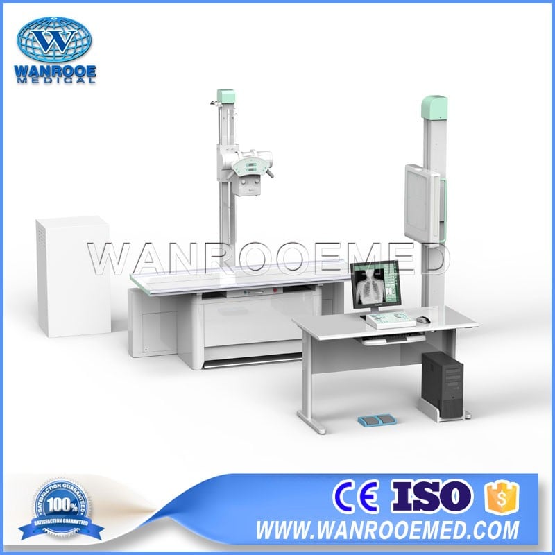
PLD3600 Hospital Digital Radiography System Fixed X Ray Machine is applied to take digital radiography on every part of human body, such as head, limbs, chest, limbus and abdomen and etc.
PLD3600 Digital Radiography System Fixed X Ray Machine Features:
1. 220V low power supply, convenient installment
2. High cost-effective FPD DR, suitable for primary hospital to realize digitalization.
3. The floating table with bucky stand can meet the photographic requirements of different standing and lying positions. The bed floating and electromagnetic lock design makes it convenient for the accurate position of the lying patient, operation is much more convenient and flexible.
4. Potable flat panel detector can take radiography for patient on wheel chair or stretcher.
5. The world leading Toshiba 14”*17”OEM digital flat Panel detector can help you get the high-definition images.
6. The leading domestic high power compact high frequency X-ray generator and high frequency power inverter makes the machine much more compact and more convenient without the extra high-voltage generator and cable.
7. The application of the KV and MA digital closed-loop control technology and the real time control of the microprocessor ensure the accuracy and repeatability of the dose.
8. The multiple automatic protection features and fault tips ensure safety during the operation process.
9. It also can be made one kind of vehicle-mounted model. Mobile medical examination and emergency use!

Item | Content | Technical Parameter | |
Power Supply | Voltage | 220V | |
Frequency | 50Hz | ||
Capacity | ≥40kVA | ||
internal resistor | ≤0.15Ω | ||
Generator | Power Output | 32KW | |
Inverter Frequency | 30 KHz | ||
Photography | Tube Voltage | 40kV—150kV | |
Tube Current | 10mA—320mA | ||
Exposure time | 1.0ms—6300ms | ||
Digital Controlled X-ray Tube | Tube Focus | 1.2mm /0.6mm (Large/Small Focus) | |
Input Power | Large/ Small focus: 40kW/ 20kW | ||
Thermal capacity | 110KJ | ||
Rotary anode speed | 2800rpm | ||
Radiography Table | Table longitudinal movement | ≥900mm | |
Table transverse movement | ≥220mm | ||
Pillar movement along table | ≥1300mm | ||
Detecting holder movement | ≥500mm | ||
Tube assembly up-down movement | 500mm-1280mm | ||
Collimator | Manual Multi-leaf | ||
Table use fixed grid | Grid density: 103L/INCH Grid ratio: 10:1 SID: 120cm Stationary type: 15″×18″ | ||
Bucky Stand | Radio-graphic device movement along pillar | ≥1300mm | |
Cassette SID | 450- 1780mm | ||
Fixed grid | Grid density 103L/INCH Grid ratio: 10:1 SID: 180cm Size: 15″×18″ | ||
Flat Panel Detector | Vision size | 358mm(H)×430 mm(V) |
Pixel Matrix | 2560(H)×3072(V) | |
Pixel Pitch | 139 μm | |
Imaging time | less than 10s | |
Limiting resolution | 3.5 lp/mm typ | |
A / D transform | 14 bit | |
Energy range | 40 - 150 kVp | |
Max input dose (low transmission gain) | 4 mR / frame | |
Date out | A 16-bit digital output Ethernet(1000BASE - T) | |
Command control | Ethernet(1000BASE - T) | |
Power input | DC 24V 4.8A |
Work Station | |
CPU System | Brand: DELL OPTIPLEX 7010 commercial use Processor: Core™ i5-3570 processor RAM: 4GB DDR3 Hardware: 500GB SATA (7200 rpm) CD-driver: suit MT’s 16X variable-speed DVD + / - RW With functions of double written Network Card: Broadcom NetXtreme 10/100/1000 PCIe Gigabit LAN GNTB-A High display card: 1GB AMD RADEON HD 7570,FH,with DVI-VGA Adapter Slop: PCIE seriel port and parallel port, all height, MT |
Color LCD Monitor | 19 inches 1M LED backing light LCD display revolution1280x1024, 5:4screen dot pitch: 0.294mm contrast ratio: 1000:1 brightness: 250cd/㎡ Grey feedback time: 5ms visual Angle: 160/170° |
Monochrome Monitor(Optional) | 1280*1024,19inches brightness 1000cd/m2 Contrast Ratio 900:1 |
Work Station Software | Basic operation: Change control console password, edit ID, acquisite images. |
Additional operation: Add new check, edit present checking info, add new position, change image aquisition order, multi-checking agreement chance, manual adjustment of exposure parameters, automatic exposure control mode, focus choice, patient body-type choice, tube capacity check, ESA curve choice, image cutting, note added (sent to DICOM workstation), mark on images, rotate or overturn, full-size image observation, check patient info and dose info, accept or refuse images. | |
Image management: Change order, patient basic info editing, inquiry history images, resend history images, re-print history images, check images mark info, review history images, manage refused images, space reclaimed, image protection, and manual image deletion etc. | |
System management: ID edition, change ID password, ED refrigeration set, statistics info checking, detector calibration, equipment control, output order management, image measurement. |
Strive to Improve Medical Conditions, Reduce Health Care Costs
© 2018-2020 Jiangsu Rooe Medical Technology Co., Ltd. All rights reserved. Site Map